The pervasive challenge of Osteoarthritis (OA), particularly affecting the knee cartilage, represents a leading cause of disability globally [3]. As a chronic, degenerative joint disease, OA involves the breakdown of articular cartilage.
This is the smooth, non-innervated, and avascular tissue that lines the ends of bones [4]. Because this tissue lacks a direct blood supply, its inherent capacity for self-repair is severely limited. In turn, this can lead to progressive joint failure [4].
Existing clinical treatments, which often include non-steroidal anti-inflammatory drugs (NSAIDs) and eventually joint replacement surgery, primarily address symptoms or mechanical failure rather than the fundamental biological pathology [3].
The scientific community is increasingly focused on developing Disease-Modifying Osteoarthritis Drugs (DMOADs). These agents are capable of altering the progression of the disease [5].
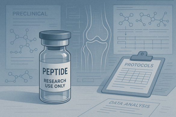
Within this specialized field, Cartalax (Ala-Glu-Asp), an ultrashort bioregulator peptide, has emerged as a subject of significant research interest. This research focus is part of the broader evidence base around cartalax peptide and cartilage-targeted bioregulation.
As a synthetic, tissue-specific agent, Cartalax is hypothesized to bypass traditional systemic signaling. It acts directly at the genetic level within the target cells: the chondrocytes [1, 2].
This comprehensive analysis details the research basis for Cartalax in OA and knee cartilage repair. For the overarching joint-focused framework, see Cartalax peptide for joint recovery.
It explores its proposed gene-regulatory mechanism, the challenges of therapeutic delivery, and the stringent protocols necessary for its study in translational orthopedic research [5].
The Pathobiology of Knee Cartilage Failure
Effective therapeutic intervention requires a deep understanding of the molecular pathology driving knee cartilage degeneration. This process is far more complex than simple mechanical erosion. It is a genetically and biochemically driven cellular catastrophe [1].
Chondrocyte Phenotypic Drift and Catabolism
The core failure in OA is the loss of the stable, quiescent phenotype of the chondrocyte. This is the sole cell type responsible for maintaining cartilage structure [1].
- Hypertrophic Differentiation: Under stress, injury, and age-related stimuli, chondrocytes undergo a pathological process called hypertrophic differentiation [1]. This transition mirrors the process by which cartilage is naturally replaced by bone during development. However, its occurrence in adult articular cartilage is destructive. The cells begin expressing markers associated with bone formation (e.g., Type X Collagen) and initiate calcification of the matrix. They fundamentally change the tissue’s mechanical properties from resilient to brittle [1].
- Destructive Enzyme Overexpression: The hypertrophic shift activates the production of potent catabolic enzymes, predominantly the Matrix Metalloproteinases (MMPs), such as MMP-13 [1, 3]. These enzymes are responsible for cleaving Type II Collagen and aggrecan. These are two key structural proteins of the cartilage matrix [1]. The balance between synthesis and degradation is lost. This leads to a net loss of matrix volume and, eventually, full-thickness lesions [4].
The Role of Senescence and Inflammation
The progression of OA is also intimately tied to cellular aging and a chronic inflammatory state [2].
- Chondrocyte Senescence: With age and cumulative damage, chondrocytes enter a state of irreversible cellular senescence [2]. Senescent cells stop dividing but remain metabolically active, secreting a devastating cocktail of proinflammatory cytokines, chemokines, and MMPs. The latter are collectively known as the Senescence-Associated Secretory Phenotype (SASP) [1]. The SASP propagates damage to neighboring healthy chondrocytes and other joint tissues, thus accelerating the cycle of degeneration [1]. For age-related lower body issues, review Cartalax For Hip Joint Health & Mobility In Aging.
- Inflammatory Signaling: Proinflammatory cytokines, particularly Interleukin-1 beta (IL-1beta) and Tumor Necrosis Factor-alpha (TNF-alpha), are abundant in the OA synovial fluid [3]. These cytokines bind to receptors on the chondrocyte surface. For related spinal applications, see Cartalax For Back Pain & Disc Degeneration: User Reports & Evidence. They trigger signaling cascades (like the NF-kB pathway) that directly upregulate MMP gene expression, perpetuating the catabolic environment and hindering any attempts at repair [3].
The challenge for any DMOAD is to target this complex pathology by suppressing catabolism, inhibiting senescence, and restoring anabolic function. These are all within the constraints of the challenging knee joint environment [5].
Cartalax: Mechanism of Action and Tissue Specificity
Cartalax (Ala-Glu-Asp) belongs to a class of peptides known as cytomedins. These short regulatory peptides were developed in Russian research, hypothesized to regulate cell- and tissue-specific functions [2]. Its power lies not in widespread signaling, but in its precise, targeted interaction with the chondrocyte’s internal regulatory machinery.
The Intracellular Advantage: Penetration and Transport
Unlike large protein therapeutics that must bind to cell surface receptors, the ultrashort nature of Cartalax, a tripeptide, provides a fundamental intracellular advantage [2].
- Size and Physicochemistry: The low molecular weight and optimized charge distribution of the Ala-Glu-Asp sequence allow it to potentially cross the cell membrane [2]. This process is hypothesized to be mediated by the cell’s natural uptake systems, specifically the Proton-coupled Oligopeptide Transporters (POT family). Examples include PEPT1 and PEPT2, or L-type Amino Acid Transporters (LATs) [2]. These transporters are designed to internalize small oligopeptides for nutritional purposes. Cartalax is believed to exploit this existing system [2].
- Bypassing the Receptor Hurdle: This transporter-mediated uptake is crucial. It allows the peptide to accumulate within the cytoplasm and nucleus, directly reaching the site of gene regulation. It also avoids the problem of widespread pleiotropy (non-specific effects across multiple tissues) that results from binding to common, ubiquitous cell surface receptors [2, 3].
Gene Regulation and Phenotype Normalization
Cartalax is proposed to function as an epigenetic bioregulator. A deeper mechanistic breakdown is covered in what Cartalax peptide is. It subtly influences the transcriptional environment of the chondrocyte [2].
- Anabolic Gene Activation: Inside the nucleus, the peptide is hypothesized to interact with chromatin or transcription factors [2]. The primary goal is to upregulate anabolic gene expression. It specifically supports the synthesis of Type II Collagen and aggrecan [1]. This is the essence of promoting repair, encouraging the cell to lay down the correct, functional matrix components [4].
- Anti-Senescence and Anti-Hypertrophy: Research suggests that short regulatory peptides can modulate markers associated with cellular aging [1]. For Cartalax, this means potentially helping the chondrocyte resist the senescent phenotype and preventing the pathological shift toward hypertrophic differentiation that drives OA [1]. By influencing gene expression, it attempts to “reprogram” the stressed or aged chondrocyte back to a stable, matrix-producing state [2]. The potential outcomes of this reprogramming are summarized in 5 Cartalax Peptide Benefits You Need To Know.
This tissue-specific and gene-regulatory mechanism is what differentiates Cartalax research from approaches using broad-spectrum anti-inflammatories or generalized growth factor signaling [3].
Research Protocols and Translational Challenges
The specialized mechanism of Cartalax dictates highly specific research and clinical protocols. It primarily concerns its delivery and the measurement of efficacy.
The Challenge of Delivery in the Knee Joint
The avascular nature of cartilage and the dynamic environment of the knee joint pose the greatest pharmacokinetic challenge for peptide therapeutics [4].
Clearance and Half-Life
If administered systemically (e.g., orally or subcutaneously), ultrashort peptides are subject to rapid systemic degradation by blood proteases and renal clearance [3]. Even if administered via intra-articular (IA) injection, simple aqueous solutions of soluble peptides are rapidly cleared from the synovial fluid, often within hours [4].
This short half-life makes it difficult to maintain the concentration above the Minimum Effective Concentration (MEC) long enough to induce a durable genetic or regenerative effect [4]. Timelines for recovery are detailed in Cartalax For Post-Injury Cartilage Repair: Timelines & Markers. This is why research protocols emphasize precise Cartalax peptide dosage and repeatable administration timing.
Formulation Imperatives
Consequently, translational protocols must incorporate advanced drug delivery systems [4, 6]. Researchers are exploring methods to functionalize biomaterials or encapsulate the peptide:
- Injectable Hydrogels: Integrating Cartalax into injectable hydrogels (such as those based on hyaluronic acid or dextran) that are designed to gradually degrade in situ offers a way to achieve sustained local release of the peptide over days or weeks [4].
- Scaffold Functionalization: For complex cartilage defects, the peptide sequence can be covalently linked to the polymer backbone of tissue engineering scaffolds. This ensures the bioactive molecule is tethered to the defect site, constantly signaling the newly recruited or implanted cells (e.g., Mesenchymal Stem Cells or MSCs) to differentiate specifically into stable chondrocytes [4, 6].
Standardized Clinical Trial Endpoints
Any therapeutic agent seeking DMOAD status must demonstrate efficacy using rigorous, objective metrics recognized by regulatory bodies [5]. Current clinical trial designs for OA peptides exemplify these standards [5].
Primary Clinical Endpoints
While pain and function remain critical, DMOAD research emphasizes the use of validated patient-reported outcome measures (PROMs), such as the Western Ontario and McMaster Universities Arthritis Index (WOMAC). These can help track changes in pain, stiffness, and physical function [5]. Other instruments include the Knee Injury and Osteoarthritis Outcome Score (KOOS) [5].
Structural Endpoints (Imaging)
To prove disease modification, not just symptom relief, trials must demonstrate a structural change [5]. This typically involves:
- Quantitative Magnetic Resonance Imaging (MRI): Used to measure changes in cartilage thickness, volume, and composition (e.g., T2 mapping provides insight into water and collagen orientation) [5]
- Radiographic Progression: Measuring the reduction in the rate of Joint Space Narrowing (JSN). Although, this marker is slow and requires long-term follow-up [5].
- Biochemical Endpoints (Biomarkers): Blood or synovial fluid samples are analyzed for changes in biomarkers of cartilage turnover [5]. Success for Cartalax would be indicated by a decrease in catabolic markers (e.g., fragments of Type II Collagen breakdown, like CTX-II) and an increase in anabolic markers (e.g., pro-collagen peptides). This validates the proposed gene-regulatory mechanism in vivo [5].
Rigorous Handling and Research Integrity
The study of ultrashort peptides requires meticulous attention to laboratory and pre-clinical protocols. These can help ensure the integrity of the active molecule [6].
- Aseptic Reconstitution: Cartalax is typically supplied in a lyophilized (freeze-dried) powder form to ensure maximum stability [6]. Step-by-step lab handling is outlined in the Cartalax peptide reconstitution guide. The reconstitution process must be performed under strict aseptic conditions, using a sterile solvent (often Bacteriostatic Water for Injection, BWFI) [6]. A critical step is sterile filtration (e.g., through a 0.2 micrometer filter) of the reconstituted solution to remove any particulates or microbial contaminants that could introduce proteases capable of degrading the peptide [6]. For ensuring initial quality, consult Cartalax Purity Guide: Testing Labs & Vendor Red Flags 2026.
- Aliquoting and Storage: Due to the risk of degradation from repeated exposure to moisture and temperature fluctuations, the stock solution must be immediately divided into small, single-use aliquots. It should also be stored at deep-freeze temperatures (e.g., minus 20 or lower) [6]. This practice prevents damage from freeze-thaw cycles. Failing to properly store the solution can induce chemical degradation (such as hydrolysis and deamidation) and physical aggregation. Common handling errors are outlined in Beginner Mistakes With Cartalax: Common Pitfalls In Research Protocols. On the other hand, proper storage can ensure the biological activity is preserved for the entire duration of the research or clinical protocol [6].
The Research Context: Cartalax Versus Other Biologics
The utility of Cartalax must be understood in comparison to other agents currently investigated for OA. This highlights the advantages of its ultrashort, tissue-specific nature.
Contrast with Systemic Growth Factors
Many therapeutic approaches for cartilage involve the delivery of large growth factor. This includes Insulin-like Growth Factor 1 (IGF-1) or Transforming Growth Factor-beta (TGF-beta) [4].
- Pleiotropy Risk: While potent, growth factors are naturally systemic and target receptors present on virtually all cell types [3]. Injecting a high concentration of a generic growth factor into the knee risks pleiotropy. This can lead to undesirable off-target effects, like the proliferation of synovial tissue or the promotion of bone spur growth in the surrounding bone, which is detrimental to joint function [4].
- Specificity of Cartalax: Cartalax’s proposed mechanism of action, targeting gene transcription within the chondrocyte, is significantly more focused [2]. By acting on the fundamental cellular decision-making process, it aims for a normalization of the cell phenotype without the excessive proliferation signals characteristic of broad-spectrum growth factors [1].
Contrast with Long-Chain Peptides
The field of peptide therapeutics includes longer sequences (e.g., 15-30 amino acids). These are often designed to mimic or block the activity of a natural protein [3].
- Immunogenicity: The risk of an immune response (immunogenicity) increases with the length and complexity of the peptide [3]. The ultrashort, natural sequence of Cartalax minimizes this risk. For a review of associated risks, see Cartalax Side Effects: Potential Complications Of This Peptide. Thus, it makes it an attractive candidate for long-term chronic treatment [3].
- Metabolic Simplicity: As a tripeptide of common amino acids (Ala, Glu, Asp), the metabolic fate of Cartalax is highly predictable. It is broken down into natural amino acid components that are readily utilized or excreted [3]. This clean metabolic profile is a significant safety advantage in the highly regulated pharmaceutical environment [3].
Integration into Future Therapies
The most promising future protocols involve integrating the high-specificity signaling of Cartalax with sophisticated delivery platforms and cellular therapies [4, 5].
- Biomimetic Scaffolds: Cartalax could be tethered to next-generation biomimetic scaffolds (e.g., those with a triple helix conformation mimicking collagen) to create an environment that is both structurally supportive and biologically instructive [4]. This combination ensures that the defect is mechanically filled. For upper body trauma, explore Cartalax For Shoulder & Rotator Cuff Injuries. Meanwhile, the cells within the scaffold are constantly signaled to adopt the desired chondrogenic phenotype [4].
- Combinational Strategies: Research is exploring combinations of regenerative agents [5]. For instance, combining the gene-regulatory signal of Cartalax with the anti-inflammatory and cellular scaffolding properties of Platelet-Rich Plasma (PRP) could lead to a synergistic effect. This addresses both the catabolic, inflammatory environment and the need for new matrix synthesis [5].
Conclusion: The Future of DMOAD Research
The development of effective therapies for knee OA hinges on overcoming the limitations of current treatments and designing agents that penetrate the joint’s hostile environment to restore cellular function. Cartalax (Ala-Glu-Asp), as a tissue-specific ultrashort peptide bioregulator, represents a critical research strategy: leveraging precise, intracellular gene-regulatory mechanisms to stabilize the chondrocyte phenotype, suppress catabolism, and promote the synthesis of Type II Collagen [1, 2].
While its full clinical potential is still being elucidated through preclinical models, the research protocols being developed, focused on advanced sustained delivery systems and rigorous structural and biochemical endpoint measurement (WOMAC, MRI, CTX-II), reflect the high scientific standard required to validate a true DMOAD [5].
For practical expectations around timelines, see how long Cartalax takes to show effects in research observations. The study of Cartalax offers valuable insights into the future of regenerative medicine. Specificity at the molecular level is the key to achieving sustained joint recovery and combating the debilitating effects of osteoarthritis.
For the main overview, see Cartalax Peptide: The Ultimate Guide For 2025.
Citations
[1] Chondrocyte Homeostasis and Differentiation: Transcriptional Control and Signaling in Healthy and Osteoarthritic Conditions – MDPI. https://www.mdpi.com/2075-1729/13/7/1460
[2] Transport of Biologically Active Ultrashort Peptides Using POT and LAT Carriers – PMC. https://pmc.ncbi.nlm.nih.gov/articles/PMC9323678/
[3] Exploring the Potential of Bioactive Peptides: From Natural Sources to Therapeutics – NIH. https://pmc.ncbi.nlm.nih.gov/articles/PMC10855437/
[4] The state of cartilage regeneration: current and future technologies – PMC. https://www.ncbi.nlm.nih.gov/pmc/articles/PMC4596193/
[5] The Current Status of Clinical Trials on Biologics for Cartilage Repair and Osteoarthritis Treatment: An Analysis of ClinicalTrials.gov Data – clinicaltrials.gov. https://pubmed.ncbi.nlm.nih.gov/35546280/
[6] Synthesis, Characterization and Evaluation of Peptide Nanostructures for Biomedical Applications
– PMC. https://pmc.ncbi.nlm.nih.gov/articles/PMC8348434/